Transthoracic Echocardiogram (TTE): Procedure, Uses, & Cost
Updated: October 14, 2024
Introduction

Want to know what goes on internally when you experience chest pains, coronary heart palpitations or dyspnea? There’s a painless method; a Transthoracic Echocardiogram (TTE) which could show the reason. We will expound significantly on TTE, such as what it can detect, the mode of action, the desired and undesired outcomes and even the cost of the procedure.
Why Do I Need a Transthoracic Echocardiogram (TTE)?
Have you ever experienced chest pain, difficult breathing, or irregular heartbeat?
This may shock you and make you wonder what can be wrong.
No need to worry anymore. There’s a painless method that would detect the cause. A US transthoracic echocardiogram (TTE) is a powerful tool that can be used to detect the cause.
So, how does it work? Let’s dive deeper and discover a few reasons why your medical doctor would possibly advice on this:
Uncovering Hidden Issues.
Chest pain, shortness of breath, or that feeling like your heart’s doing a faucet dance . Those signs and symptoms could be an indication that something is happening with your coronary heart. A TTE allows you to visualise the culprit, whether it is narrowed arteries, funky valves, or a weak heart muscle.
Keeping Your Valves in Check
A TTE can assess their features, figuring out abnormalities like stenosis (narrowing) or regurgitation (leakage). Early detection is important in monitoring the condition and preventing complications down the line.
Diagnosing Heart Conditions

A TTE can be a lifesaver while diagnosing heart failure [failure of heart muscle to pump blood] and cardiomyopathy .Knowing the cause can help the doctor to come up with a treatment method.
Even if you were born healthy, a TTE can detect hidden congenital heart defects, meaning abnormalities present since birth. Early diagnosis allows for proper management and improves your long term health outcomes.
Pregnancy
A TTE can also be done to visualise the fetal heart in the womb in high risk pregnancies to screen for any cardiac abnormality which may require early treatment.
Inquire from your doctor whether a TTE is right for you.
Remember, early detection is prime!
Reasons for a Transthoracic Echocardiogram
Conditions Diagnosed by TTE
Feeling a little tightness or fluttering? . Let’s discover what a TTE can display.
- Heart Valve Disease
Think of your coronary heart valves as doorways. TTE lets medical doctors see if those valves are narrowed (stenosis), leaky (regurgitation), or have abnormal attachments, all of which can have an effect on blood flow..
- Heart Failure
A weakened heart can not pump blood efficiently. TTE measures your heart chamber size and pumping efficiency to diagnose heart failure and determine its severity.
- Myocardial Infarction (Heart Attack]
The coronary arterial blockage causing heart attack can be clearly visualised by a TTE as well as the entire heart strcture.It can also asses the general function of the heart post heart attack
- Congenital Heart Defects
Born with a coronary heart murmur? TTE performs a crucial role in detecting structural abnormalities like abnormal blood vessels, holes inside the coronary heart wall or narrowed valves.
- Pericardial Effusion
TTE can detect over accumulation of pericardial fluid around the heart. This complication can be fatal if not diagnosed early.
Early detection is fundamental to handling coronary heart fitness. If you’re worried about your heart, communicate to your physician extensively whether or not a TTE is proper for you.
Transthoracic Echocardiogram Procedure
How Do You Prepare for a TTE?
A TTE is a simple procedure. In most cases, there’s no special prep wanted. However, your doctor may advocate skipping that heavy lunch beforehand since a full belly can cause discomfort.. Loose garments are your friend for this one. Think cozy pajamas or a comfortable blouse, something that allows access to your chest.
Inform your physician if you are allergic to any medications, especially the ultrasound gel to be used.. It’s additionally beneficial to mention any medicines that you are presently taking. While a TTE is a painless technique, you may feel slight discomfoort from lying on the table for a while. The technician will check on you continuously to ensure you are comfortable.
The Big Day, What Happens During a TTE?
Relax, It’s Painless
The complete procedure is quite easy and simply takes 30-45 mins. During this procedure, you will be witnessing the problematic performance of your heart valves, chambers, and blood flow. Here’s how it is going:
The TTE is highly quick, usually lasting around 30-45 mins. You’ll be positioned comfortably on your left side . There’s no need to worry about complicated prep. Generally, a light snack beforehand is all it takes. The technician will apply a groovy gel on your chest. This unique gel enables the sound waves to tour smoothly, allowing them to capture an accurate picture of your heart’s inner workings.
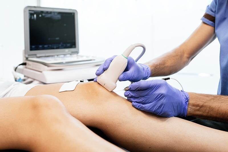
The transducer emits sound waves that bounce back from your coronary heart, like an echo coming back from the walls of a hidden chamber. The technician will expertly maneuver the transducer throughout specific regions of your chest. It’s like capturing the live performance from diverse angle:
- Short axis view: This view offers a cross-sectional picture, like peering through a window, revealing the chambers and valves in detail.
- Long axis view: Imagine a full body image of your coronary heart. This view gives a breathtaking look, showcasing the overall structure and motion of your coronary heart chambers..
- Apical views: Think of these views as zooming in at the tip of your heart. They provide magnified pictures of the valves, ensuring no hidden details go unnoticed..
To actually understand your coronary heart’s symphony, TTE makes use of Doppler generation. By studying the direction and speed of blood flow. Doppler can identify issues like leaky valves or narrowed pathways, making sure your coronary heart’s rhythm remains regular.
After the photo acquisition and Doppler evaluations are complete, you will have detailed information of your heart’s health. This statistics can be checked by your doctor, helping them to diagnose any existing issues and make sure your heart maintains its excellent overall performance for years to come.
After the Procedure
Following your us transthoracic echocardiogram (TTE), the technician will carefully wipe away the gel and you will be free to return on your day. But what about the pictures? The sonographer will evaluate them intently and send them to a heart specialist for his or her expert interpretation.
The time-frame for receiving your results can range depending on your health practitioner’s schedule, however in many cases, you would expect to hear from your doctor within a few days with a detailed clarification . Imagine the relief of having those answers!
Transthoracic Echocardiogram (TTE) vs. Transesophageal Echocardiogram (TEE)
Hold on, there’s more than one type of echo?
It can be confusing with these kinds of medical abbreviations! There are commonly two types of echocardiograms used to assess your heart: US Transthoracic (TTE) and Transesophageal (TEE). Both use sound waves to create photos, but how they get the images is quite different.
TTE
TTE is mostly used during echo. It’s absolutely painless. A technician virtually glides a handheld device throughout your chest to capture photos of your heart. Think of it like taking a photo of your private home from the sidewalk. Although it gives a good general view the smaller details are blurring
TEE
TEE takes matters a step further. A thin, flexible probe is inserted down your esophagus (the tube that connects your mouth to your belly). Since it’s located very close to your heart, TEE offers a more detailed photograph. Basically it zooms into your heart and magnifies the small structures clearly. Because the probe is inserted it requires sedation to ensure comfort.
Here’s the bottom line:
Both TTE and TEE are valuable gear for diagnosing coronary heart troubles. Your medical doctor will advise on the best option based on your condition..
Okay, so TTE sounds pretty convenient. But when might a doctor opt for a TEE as an alternative?
While TTE is the most preferred for majority of heart checkups, TEE shines in some specific situations:
Improved visualization of specific structures
This close-up view is mainly useful for examining:
- The back of heart: Structures like the posterior wall, mitral valve, and aortic valve are all tucked away within the lower back. TEE offers a clearer image of these regions.
- Unclear TTE photos: Sometimes, factors like lung troubles, more weight, or a wiggly chest wall can make TTE photos blurry. TEE bypasses those limitations for a sharper view.
Suspected valve abnormalities
TEE’s detailed view allows doctors to investigate valve features, specifically the mitral and aortic valves. This can be essential for figuring out issues like valve prolapse or leakage (regurgitation).
Evaluation of tumors near the coronary heart
If there may be a mass or tumor lurking close to your heart, TEE can offer a precise picture. This facilitates doctors to better recognize the situation and determine the next step.
So, which one will you get?
The selection of whether or not to undergo a TTE or TEE may be made by your doctor based on your specific condition. TTE is commonly the first line preference due to its non-invasive nature and simplicity of use. However, in case your physician needs a greater distinct evaluation of particular structures or the TTE images are blurred, TEE is probably endorsed.
Transthoracic Echocardiogram Results

Understanding medical jargon may be tricky, but here is a breakdown of these cryptic details..
How is TTE Results Interpreted?
During a TTE, the dimensions and thickness of these chambers and valves are precisely measured. These measurements are then compared to normal values to detect any abnormalities. A moderate difference might be okay, however a full size deviation should be further investigated.
Ejection Fraction
This is an important value that reflects how efficiently your heart pumps blood with each beat. A regular EF usually falls in the range of 55% to 70%. A lower EF, however, might suggest a weakened heart, prompting further research. Now, this doesn’t always suggest there’s prime trouble, but it does warrant further research along with your physician.
Blood Flow: Keeping Things Running Smoothly.
TTE uses of Doppler principle to evaluate the direction and speed of this blood flow. Normal flow resembles smooth, one-manner traffic. However, abnormal flows include turbulence or disruption, indicate problems like valve leakage or narrowing. These problems are like traffic jams in your heart, hindering its efficient operation.
A heart specialist is best equipped to interpret the whole image considering your measurements, EF, blood flow patterns, and medical history. They can provide an explanation for what the results imply for you and advise on the next steps for optimal heart health.
What Happens After Receiving Abnormal Results?
Uh oh! TTE results not looking quite right?
Listen, getting strange results on a test may be scary. You are probably wondering what it means and what takes place next.
Don’t panic!
While atypical outcomes can indicate an underlying difficulty, it is crucial to recognize what they suggest inside the context of your specific health.
Here’s what to expect:
Your medical doctor will walk you through the findings. They’ll explain what the abnormalities mean and how they are probably affecting your cardiac function..
Depending on the findings additional exams might be needed. This should consist of things like a stress echo or a cardiac cath.
In some instances, a specialist is preferred such as a cardiothoracic surgeon to further view the results and provide treatment options.
The good news?
Treatment plans vary depending on the analysis, it may include medicines, lifestyle changes, minimally invasive procedures, or even open heart surgical procedure. Your medical doctor will collaborate with you to create a customized plan to cover your particular needs and get your heart health headed in the right direction.
Empower yourself by speaking to your health practitioner and understanding your results. Early detection and treatment are key to managing any heart conditions.
Still have questions?
Don’t hesitate to call your physician for an explanation. They’re there to guide you through this procedure.
Transthoracic Echocardiogram Cost
Alright, so understanding your TTE results is important, but what about the value?
We realize money matters, so let’s speak on what to expect on the subject of the invoice in your TTE.
Here’s the element, the cost can range relying on some factors:
Insurance
Having medical insurance is a big benefit. Your plan negotiates decrease charges with hospitals and clinics. But how much you certainly pay depends on your specific plan details including deductible, copay, and coinsurance. It’s like a decoder ring you want to crack the price.
Facility fees: Hospital vs. Clinic?
Hospitals regularly have better charges in comparison to outpatient clinics. Think fancy motel room vs. Cozy bed and breakfast each offers a bed but the charge reflects the pricing.
Location.
Costs can fluctuate depending on where you live. Areas with high cost of living may suggest pricier medical services, inclusive of TTEs.
So, how much does it cost?
There is no standard fee. However the cost ranges approximately between $1,000 to over $3,000. Your real price might be higher or lower.
Before scheduling your TTE, call your insurance organization and these questions:
- Are there in network providers for TTEs
- Is TTE included in my plan?
- Do I need a referral?
- What’s my deductible for this?
- What are my copay or coinsurance charges?
Conclusion
Don’t wait to prioritize your heart fitness! Schedule a session with your health practitioner to see if a TTE is proper for you. Early detection is prime, so take control of your wellbeing and keep your coronary heart happy and healthy. Would you be curious to know more about healthy heart habits? Share your questions and feedback beneath!
Sources
- Attia, Z.I., Harmon, D.M., Behr, E.R. and Friedman, P.A. (2021). Application of artificial intelligence to the electrocardiogram. European Heart Journal, 42(46), pp.4717–4730.
- Reardon, R.F., Chinn, E., Plummer, D., Laudenbach, A., Andie Rowland Fisher, Smoot, W., Lee, D., Novik, J., Wagner, B., Kaczmarczyk, C., Moore, J., Thompson, E., Tschautscher, C., Dunphy, T., Pahl, T., Puskarich, M.A. and Miner, J.R. (2021). Feasibility, utility, and safety of fully incorporating transesophageal echocardiography into emergency medicine practice.Academic emergency medicine, 29(3), pp.334–343.
- Boissier, F., Bagate, F. and Mekontso Dessap, A. (2020). Hemodynamic monitoring using trans esophageal echocardiography in patients with shock. Annals of Translational Medicine, [online] 8(12), p.791.
- Patel, K.M., Desai, R.G., Trivedi, K., Neuburger, P.J., Krishnan, S. and Potestio, C.P. (2022). Complications of Transesophageal Echocardiography: A Review of Injuries, Risk Factors, and Management. Journal of Cardiothoracic and Vascular Anesthesia, 36(8), pp.3292–3302.
- Freitas-Ferraz, A.B., Bernier, M., Vaillancourt, R., Ugalde, P.A., Nicodème, F., Paradis, J.-M., Champagne, J., O’Hara, G., Junquera, L., del Val, D., Muntané-Carol, G., O’Connor, K., Beaudoin, J. and Rodés-Cabau, J. (2020). Safety of Transesophageal Echocardiography to Guide Structural Cardiac Interventions. Journal of the American College of Cardiology, 75(25), pp.3164–3173.
- Parker, B.K., Salerno, A. and Euerle, B.D. (2018). The Use of Transesophageal Echocardiography During Cardiac Arrest Resuscitation: A Literature Review. Journal of Ultrasound in Medicine, 38(5), pp.1141–1151.
- Hatala, R., Gutman, J., Lineberry, M., Triola, M. and Pusic, M. (2018). How well is each learner learning? Validity investigation of a learning curve-based assessment approach for ECG interpretation. Advances in Health Sciences Education, 24(1), pp.45–63.
- Orihashi, K. (2019). The history of transesophageal echocardiography: the role of inspiration, innovation, and applications. Journal of Anesthesia, 34(1), pp.86–94.
- Jozwiak, M., Mercado, P., Jean-Louis Teboul, Anouar Benmalek, Giménez, J., François Dépret, Richard, C. and Monnet, X. (2019). What is the lowest change in cardiac output that transthoracic echocardiography can detect? Critical Care, 23(1).

Article by
Scott Caswell
Scott is a co-founder of PUM and an ultrasound technology expert with a passion for innovation in the medical field. Scott has dedicated his career to advancing portable ultrasound devices, making medical imaging more accessible to professionals around the globe.
When not refining ultrasound devices, he enjoys hiking, experimenting with new recipes, and exploring the latest tech gadgets. Scott is dedicated to making healthcare more accessible and efficient through cutting-edge ultrasound solutions.

Join Our Mailing List & Save!
Enter your email address below to receive exclusive promotions and discounts along with additional product information and tips
Shop Now
Leave a Reply