How to Use a Portable Ultrasound Machine
Updated: January 6, 2025
Portable ultrasound machines have transformed healthcare, offering a mobile solution for diagnostic imaging across a variety of clinical and field settings. These machines are essential in today’s fast-paced medical environments, allowing healthcare providers to perform precise and efficient imaging.
This guide will comprehensively explain how to use a portable ultrasound machine effectively while ensuring safety and accuracy.
What Is the Basic Knowledge About Portable Ultrasound Machines?
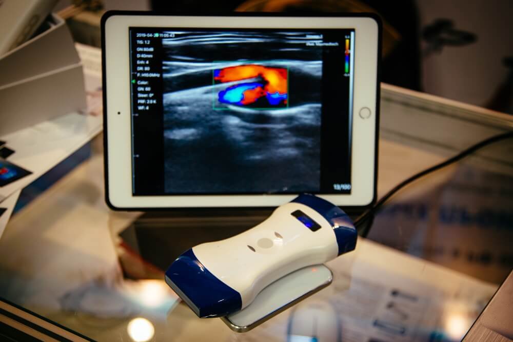
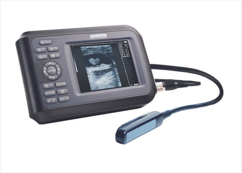
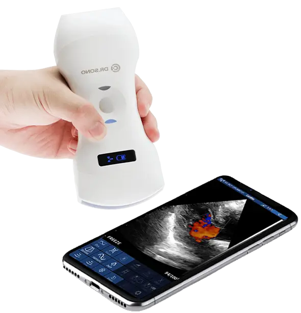
Portable ultrasound machines are compact devices that use high-frequency sound waves to produce real-time images of the body’s internal organs, tissues, and blood flow. Unlike traditional, stationary ultrasound machines, their portability makes them ideal for use in clinics, emergency rooms, ambulances, and even remote areas.
They are widely used in fields such as obstetrics, cardiology, musculoskeletal diagnosis, and even veterinary medicine. Modern portable ultrasound machines are equipped with advanced imaging features, touch-screen interfaces, and wireless connectivity for seamless operation.
Importance of Proper Use and Safety Measures
- Accurate Diagnosis: Proper usage ensures high-quality imaging, leading to more accurate diagnoses.
- Patient Safety: Misuse can cause patient discomfort or inaccurate readings, potentially leading to misdiagnosis.
- Equipment Longevity: Handling the machine correctly prevents wear and tear, ensuring a longer lifespan.
- Infection Control: Proper disinfection of probes and the machine reduces the risk of cross-contamination.
What Are the 3 Basic Components of an Ultrasound Machine?
External Equipment Used in Portable Ultrasound Machines
- Transducer (Probe)
The most critical component, responsible for sending and receiving sound waves is a transducer. Different probes (linear, convex, or phased array) are used for specific imaging purposes.
- Display Screen
The display screen lets you see real-time ultrasound images. High-resolution screens improve the ability to visualize intricate details.
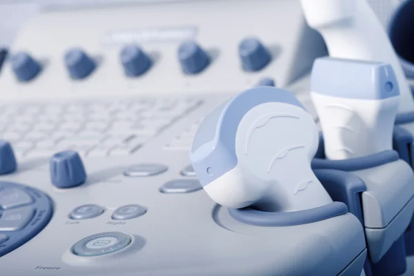
- Control Panel or Interface
It includes knobs, buttons, and touchscreen options to adjust settings such as gain, depth, and frequency for better imaging results.

Importance and Functioning of Portable Ultrasound Components
- Transducers: Different transducers cater to specific applications—linear probes for vascular and musculoskeletal imaging, curved probes for abdominal scans, and phased array probes for cardiac imaging.
- Display Screen: Ensures operators can interpret results accurately in real time, reducing the need for repeat scans.
- Control Panel: Allows fine-tuning of imaging parameters to optimize clarity and diagnostic precision.
Pre-Procedure Checklist
Before using a portable ultrasound machine, thorough preparation is crucial:
- Ensure Proper Training and Certification:
- Only certified professionals should handle ultrasound equipment.
- Proper training ensures competence in machine operation and interpretation of results.
- Familiarize Yourself with the Machine:
- Spend time exploring the machine’s interface, settings, and functionality.
- Understand the purpose of each transducer and the settings required for different imaging tasks.
- Prepare the Examination Area:
- Choose a clean, well-lit, and quiet space.
- Have all necessary supplies, like ultrasound gel, wipes, and gloves, within reach.
- Prioritize Patient Comfort and Safety:
- Provide a private environment.
- Use pillows or adjustable beds to position the patient comfortably.
- Review Patient Documents:
- Understand the patient’s medical history, allergies, and reason for the ultrasound.
- Clarify any specific areas to focus on during the scan.

Step-by-Step Procedure
1. Machine Setup and Preparation
- Switch on the machine and allow it to run self-diagnostic checks to ensure all components function properly.
- Select a probe suitable for the scan type (e.g., linear for vascular or curved for abdominal).
- Adjust settings like frequency, depth, and gain to match the imaging requirements.
- Spread a thin layer of gel on the transducer or patient’s skin to eliminate air gaps and improve sound wave transmission.
2. Patient Preparation and Positioning
- Clearly explain the process and purpose of the scan.
- Address any questions or concerns the patient may have before starting.
- Arrange the patient in the position required for the scan (e.g., supine for abdominal imaging or lateral for cardiac imaging).
- Use pillows, sheets, or other aids to make the patient comfortable.
- Regularly check on the patient during the procedure.
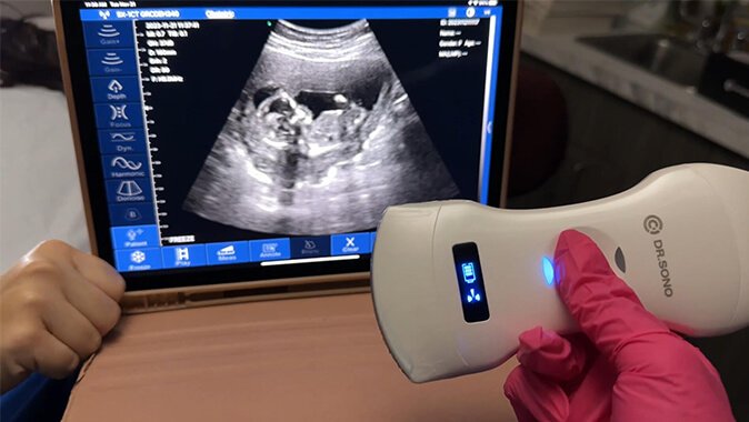
3. Image Acquisition and Optimization
- Gently press the probe against the patient’s skin.
- Modify depth, focus, and gain settings for a clearer image.
- Sweep the probe longitudinally and transversely to capture comprehensive images.
- Adjust the time-gain compensation (TGC) to enhance image sharpness in specific regions.
4. Image Interpretation and Reporting
- Identify any abnormalities, measure dimensions, and evaluate structures or blood flow patterns.
- Record key measurements, abnormalities, and observations in detail.
- Save all images securely in the machine or transfer them to a cloud storage system, adhering to privacy regulations.

Safety Measures and Troubleshooting
Safety Precautions
- Sterilize probes before and after use.
- Avoid excessive probe pressure to prevent patient discomfort.
- Regularly inspect cords and connections for electrical safety.
Troubleshooting Common Issues
- Image Quality Problems: Adjust gain and focus settings or clean the probe lens.
- Machine Malfunctions: Reboot the device or consult technical support.
- Probe Damage: Replace worn or damaged probes promptly.
Emergency Procedures
- Be prepared for patient adverse reactions (e.g., allergic reactions to gel).
- Have backup equipment ready in case of machine failure.
Tips and Best Practices
- Optimize Image Quality: Use appropriate transducers and adjust settings dynamically.
- Maintain Documentation Standards: Accurately record and store findings to avoid errors.
- Reduce Patient Discomfort: Use warm gel and communicate effectively to keep patients relaxed.
- Maintain Probes: Clean and inspect probes regularly for wear and tear.

Common Applications and Clinical Scenarios
- Musculoskeletal Imaging: Diagnose tendon, ligament, and muscle injuries.
- Cardiovascular Imaging: Evaluate heart function, and blood flow, and detect vascular conditions.
- Abdominal Imaging: Examine the liver, kidneys, pancreas, and other abdominal organs.
- Obstetric Imaging: Monitor fetal growth and assess prenatal health.
- Veterinary Imaging: Diagnose conditions in animals’ musculoskeletal, cardiovascular, and abdominal systems.
Maintenance and Quality Control
- Consistent Cleaning: Disinfect the machine and probes after every use to prevent infections.
- Routine Maintenance: Schedule regular servicing to keep the machine in optimal condition.
- Quality Checks: Conduct periodic tests to ensure imaging accuracy and equipment reliability.
Conclusion
Portable ultrasound machines are a cornerstone of modern diagnostic imaging, offering unparalleled convenience and accuracy. By mastering their usage, healthcare professionals can improve patient outcomes, streamline diagnostics, and uphold high standards of care.
Follow proper procedures, prioritize safety, and maintain the machine diligently to ensure it remains a reliable diagnostic tool for years to come.

Article by
Scott Caswell
Scott is a co-founder of PUM and an ultrasound technology expert with a passion for innovation in the medical field. Scott has dedicated his career to advancing portable ultrasound devices, making medical imaging more accessible to professionals around the globe.
When not refining ultrasound devices, he enjoys hiking, experimenting with new recipes, and exploring the latest tech gadgets. Scott is dedicated to making healthcare more accessible and efficient through cutting-edge ultrasound solutions.
Join Our Mailing List & Save!
Enter your email address below to receive exclusive promotions and discounts along with additional product information and tips
Shop Now
Leave a Reply