What Is a Pelvic Scan Looking For? Key Pelvic Ultrasound Facts to Know
Updated: August 20, 2024
A pelvic ultrasound has become an essential part of the diagnosis and treatment of the malaises that threaten pelvic health, especially among women.
There are two key reasons for this.
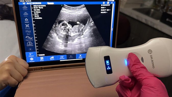
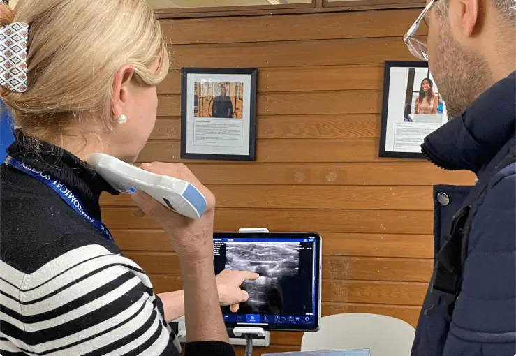
The first is the availability of ultrasound machines at the point of care, including portable handheld ultrasound scanners. As a result, pelvic sonography is always part of the routine patient assessment performed by gynecologists and obstetricians.
The second is the widespread training in pelvic ultrasound procedures and the knowledge of how to read and interpret ultrasound images and reports among specialized and general medical practitioners.
So, what is a pelvic scan looking for, and what should every woman (and man) know about pelvic ultrasound? You have all the answers in this article.
What is a Pelvic Ultrasound?
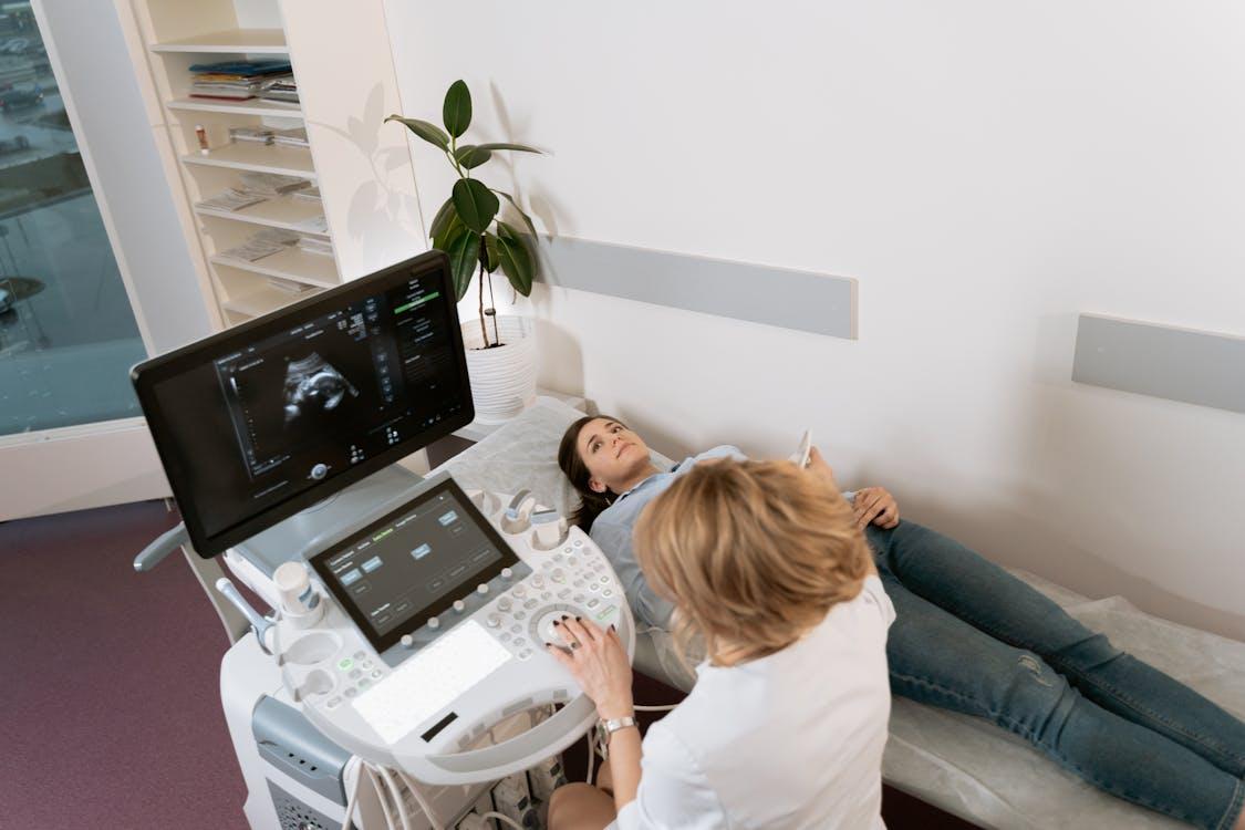
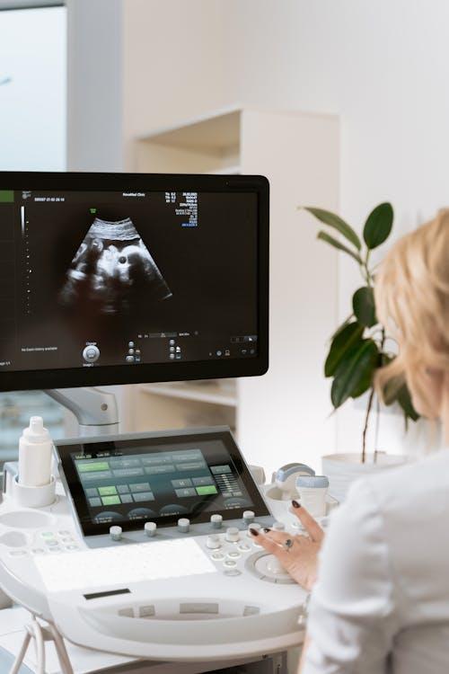
A pelvic ultrasound is a non-invasive medical exam that uses an ultrasound scanner to create real-time images of body organs in the pelvic area. This happens without the sonographer introducing an instrument into the body through the skin or a body opening.
So, how does the ultrasound create the images?
- The sonographer passes a transducer or probe connected to an ultrasound monitor on the pelvic area.
- The wand-like probe generates sound waves.
- The sound waves pass through body tissues and fluid to hit the targeted pelvic organ(s).
- The ultrasound waves, which are beyond human auditory capacity, bounce back to the probe from the organs as echo sounds.
- The ultrasound computer receives electrical signals of the echoes and converts them into images on the ultrasound monitor.
Apart from creating images, ultrasound machines with the Doppler probe mode also show the state of blood flow in the blood vessels of pelvic organs using color images.
Body organs in the pelvic area scanned with a pelvic ultrasound include:
| In Women: | In Men: |
| Uterus | Bladder |
| Cervix | Seminal vesicles |
| Ovaries | Prostate |
| Fallopian tubes | Other organs and structures of the abdomen. |
| Bladder | |
| Pouch of Douglas (rectouterine pouch) | |
| Vagina | |
| Pelvic blood vessels (Doppler ultrasound) |
You may see a pelvic ultrasound called by several other names, including gynecological ultrasound, pelvic sonogram, pelvic ultrasonogram, abdominal ultrasound, transabdominal ultrasound, and pelvic scan.
Medical personnel may also describe a pelvic ultrasound by the purpose of the procedure, like ultrasound for pelvic pain, pelvic ultrasound for fertility, ultrasound for pelvic masses, ovarian ultrasound, pelvic ultrasound for fibroids, endometrial ultrasound, and pelvic inflammatory disease ultrasound.
While the pelvic ultrasound procedure is commonly used with women, medics also conduct pelvic ultrasounds on men.
And that detail leads us to the question, what are the types of pelvic ultrasound?
What are the 3 Types of Pelvic Ultrasound
If you ask medical professionals and sonographers, they will tell you there are generally three types of pelvic ultrasound, categorized by the probing or scanning mode.
Abdominal or Transabdominal Ultrasound
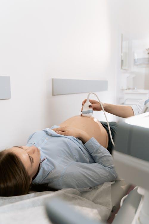
An abdominal or transabdominal ultrasound is an ultrasound mode that scans the pelvic organs by passing an ultrasound probe on the belly.
The sonographer applies a conductive gel on the patient’s belly and passes the probe to image the pelvic organs transabdominal (across or on the other side of the abdomen).
Abdominal ultrasounds are the most common form of pelvic ultrasound, also used during pelvic ultrasound pregnancy scans.
Transvaginal Ultrasound
A transvaginal ultrasound creates obstetric and pelvic images, especially of the reproductive organs, by introducing a long, thin probe into the vagina.
The probe is covered with a conductive gel and then wrapped with a latex sheath before being introduced into the vagina.
Rectal Ultrasound
A rectal ultrasound is used to image the internal side of the rectum in both women and men. However, a specialized transrectal ultrasound is used for prostate examinations in men.
Bear in mind that the male pelvic area may also be examined with abdominal ultrasound.
Medics consider two key aspects to decide on the type of pelvic ultrasound applicable in each patient case:
- The symptoms that point to the need for a pelvic ultrasound.
- The desired pelvic ultrasound results for diagnosis and treatment, or what the ultrasound is looking for.
What is a Pelvic Scan Looking For?
What a pelvic scan looks for depends on the type and purpose of the pelvic ultrasound procedure. In that light, we can break down the question of what is a pelvic scan looking for into three specific questions:
- What is a diagnostic pelvic ultrasound looking for?
- What is a pregnancy pelvic ultrasound looking for?
- What is an interventional pelvic ultrasound looking for?
Diagnostic Pelvic Ultrasound
A diagnostic pelvic ultrasound looks for malaise or abnormal tissues and structures in the pelvic organs.
In women, a diagnostic pelvis ultrasound is looking for:
- Abnormal shape, size, or position of the ovaries and uterus and the thickness of the endometrium.
- Ovarian cysts and tumors.
- Ectopic pregnancy.
- Uterine fibroids.
- Ovarian torsion.
- Pelvic inflammatory disease.
- Endometriosis.
- Polycystic ovary syndrome (PCOS).
- Pelvic organ prolapse.
In men, diagnostic pelvic ultrasound is looking for:
- Cancerous tissues in the prostate or testicular.
- Signs of scrotal and testicular infection.
- Injuries on the penis or scrotal.
- Cysts and infections in the seminal vesicles.
- Enlarged prostate or benign prostatic hypertrophy (BPH).
- Prostate volume estimate.
In both women and men, diagnostic pelvic ultrasound looks for:
- Hernias.
- Signs of bladder cancer (not a definitive cancer diagnosis).
- Kidney stones that have moved into the bladder.
- Abdominal aortic aneurysm, especially in seniors.
Using the Doppler probe mode, a diagnostic pelvic ultrasound will also look at the state of blood flow in the organs of the pelvic area.
Pregnancy Pelvic Ultrasound
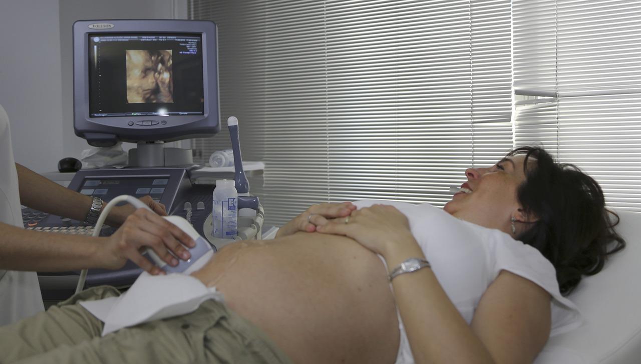
Sonographers, obstetricians, and gynecologists perform a pregnancy pelvic ultrasound to monitor the health, growth, and development of a fetus.
Specifically, a pelvic ultrasound pregnancy scan will record the fetal heartbeat, a multiple pregnancy, and abnormalities in the fetus’ head and other organs, among others.
Interventional Pelvic Ultrasound
Interventional pelvic ultrasound is mostly used to draw tissues for diagnostic purposes. For example, a specialized form of interventional pelvic ultrasound known as transperineal prostate biopsy is common in examining men for prostate cancer. A rectal ultrasound guides the biopsy needle introduced into the prostate through the area between the rectum and the scrotum.
Gynecologists and obstetricians also use interventional pelvic ultrasound to guide a needle when collecting eggs from the ovaries for IVF.
Pelvic ultrasound may also have other medical uses. For example, it can be used to confirm that an IUD (intrauterine device) is positioned safely in the uterus.
Indications for Pelvic Ultrasound?
While pregnancy pelvic ultrasounds are routine checks during the gestation period to monitor fetal growth and health, your doctor will recommend a diagnostic and interventional ultrasound if these symptoms are noticed:
- Pain in the abdominal or pelvic area.
- A swollen abdomen.
- Painful sex.
- Pain while emptying the bladder.
- Inability to conceive.
- Abnormal monthly flows or bleeding post-menopause.
- Urine leakage (urinary incontinence) or the urgency to go often.
- Urinary retention.
- Blood in the urine (hematuria).
What To Expect Before, During, and After a Pelvic Ultrasound?
A pelvic ultrasound is a safe medical exam. Apart from being non-invasive, it does not involve any radiation. Plus, it does not cause any pain, except for a slight discomfort from the probe pressing on a full bladder and the insertion of the long probe into the vagina during a transvaginal ultrasound.
That said, here’s what to expect before, during, and after a pelvic ultrasound.
Before a Pelvic Ultrasound
- Your doctor will ask you to drink water and have a full bladder before an abdominal ultrasound. You do not need to fast.
Instead, you’ll need to have an empty bladder before a transvaginal ultrasound.
For transrectal ultrasound, a bowel preparation to clear the colon is done with a suppository or enema 1-4 hours before.
- The doctor explains what the ultrasound procedure entails and allows you to ask questions or give any important information, like sensitivity or allergic reactions to latex.

- Wearing easy-to-remove clothing makes it easier to change into a gown if required or fold it up to expose your belly. Consider that traces of the transducer gel may remain on the skin, so wear clothing that won’t spoil if that is the case.
- Your doctor may give specific preparation details for your case
During a Pelvic Ultrasound
- The doctor or sonographer asks you to change into a gown and remove any jewelry or accessories that can interact with the ultrasound waves or the machine parts.
- You’ll be asked to lie supine (back down) on the exam table.
For the transvaginal ultrasound, the sonographer will also prop up your legs and feet.
For the transrectal ultrasound, you lie on the side with your knees bent. - The sonographer applies the transducer gel on your abdomen and passes the probe to examine the pelvic organs. The sonographer may invite you to look at the images.
The gel is applied to the transducer for the transvaginal ultrasound, and a latex sheath covers it. The sonographer moves the probe gently inside the vagina to find the right angle for clear imaging.
A lubricated probe is inserted into the rectum for the transrectal ultrasound and moved gently to different angles to take the needed images. - Once complete, the sonographer wipes the gel on your belly and invites you to empty your bladder and change back into your clothes.
After the Pelvic Ultrasound
You do not need to follow any special precautions after a pelvic ultrasound unless your doctor gives you specific instructions for your case.
The sonographer sends the pelvic ultrasound images and the report to your doctor to guide diagnosis and treatment.
If no malaise is found, you have a clean bill of health to take home.
Signs of a Bad Pelvic Ultrasound
A pelvic ultrasound is bad if it reports abnormalities in the pelvic organs.
Some common signs of a bad pelvic ultrasound are:
- Cysts and masses in the uterus, ovaries, or other pelvic organs. These could be indicative of cancer or other severe health conditions.
- Fluid in the pelvis due to conditions such as abnormal bleeding, ectopic pregnancy, broken ovarian cysts, and pelvic inflammatory disease.
- Thickened uterus lining, which could signal serious conditions such as endometrial hyperplasia, a precondition for endometrial cancer.
- Unclear pelvic scan images due to issues such as extreme obesity, traces of barium in the bowel following a barium swallow test, intestinal gas, and an empty or semi-full bladder. These issues make viewing of the pelvic organs inconclusive, warranting a repeat of the procedure or alternative tests.
Quick Summary
Obstetricians, gynecologists, and other medical professionals use pelvic ultrasound as a common diagnostic or interventional procedure at the point of care.
The non-invasive test creates real-time images of the pelvic organs, allowing your doctor to notice abnormal structures or monitor the growth and health of a developing fetus.
While not as common as in women, pelvic ultrasound is also used with men to examine them for prostate cancer and other ills of the pelvic organs.
A pelvic ultrasound may return positive results even though you may have the indications for a pelvic ultrasound such as pain and swelling in the pelvic area,
Talk and work with your doctor if the procedure shows signs of a bag pelvic ultrasound such as cysts and a thickened endometrium.
Top-notch Pelvic Ultrasound Imaging with Dr. Sono
At Dr. Sono, we work with individual medical practitioners and medical facilities to take pelvic ultrasound imaging to the next level. With our state-of-the-art portable diagnostic ultrasound machines, you can assure your patients quality pelvic imaging for precise diagnosis and treatment. Talk to us today for all your pelvic ultrasound solutions.
Sources
- Baylor College of Medicine: What is the Definition of Pelvic Health?
- Cleveland Clinic: Pelvic Ultrasound.
- Johns Hopkins Medicine: Pelvic Ultrasound.
- NIH: Pelvic Ultrasound.
- Corewell Health: Understanding Pelvic Ultrasound.
- Health University of Utah: Pelvic Ultrasound.
- UF Health: Transperineal Biopsy for Prostate Cancer.
- Radiopaedia: Male pelvic ultrasound (technique).

Article by
Scott Caswell
Scott is a co-founder of PUM and an ultrasound technology expert with a passion for innovation in the medical field. Scott has dedicated his career to advancing portable ultrasound devices, making medical imaging more accessible to professionals around the globe.
When not refining ultrasound devices, he enjoys hiking, experimenting with new recipes, and exploring the latest tech gadgets. Scott is dedicated to making healthcare more accessible and efficient through cutting-edge ultrasound solutions.


Join Our Mailing List & Save!
Enter your email address below to receive exclusive promotions and discounts along with additional product information and tips
Shop Now

Leave a Reply