Can a Portable Ultrasound Machine be Used to Find a Vein?
Updated: January 6, 2025
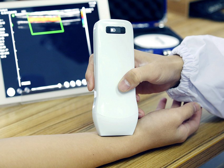
Portable ultrasound machines are compact, highly versatile devices that use high-frequency sound waves to generate real-time images of the body’s internal structures. Their mobility and ease of use have made them indispensable in various clinical settings. Do you really want to know how can a portable ultrasound machine be used to find a vein? We’ve got the answer!
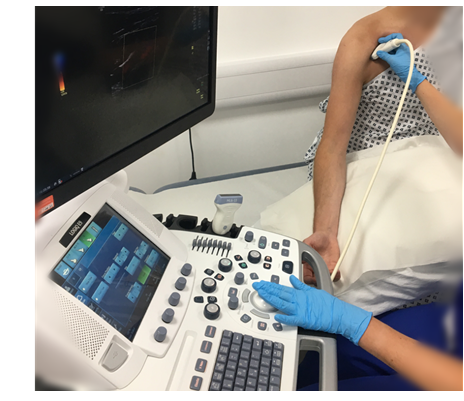
One of their emerging applications is in the precise location of veins, which is critical for procedures like intravenous (IV) cannulation, blood sampling, and central venous catheter placement. For patients with difficult venous access (DVA), finding veins can be a challenging and time-consuming process. Using ultrasound guidance can significantly improve accuracy, reduce complications, and enhance patient safety.
Can a Portable Ultrasound Machine Be Used to Find a Vein?
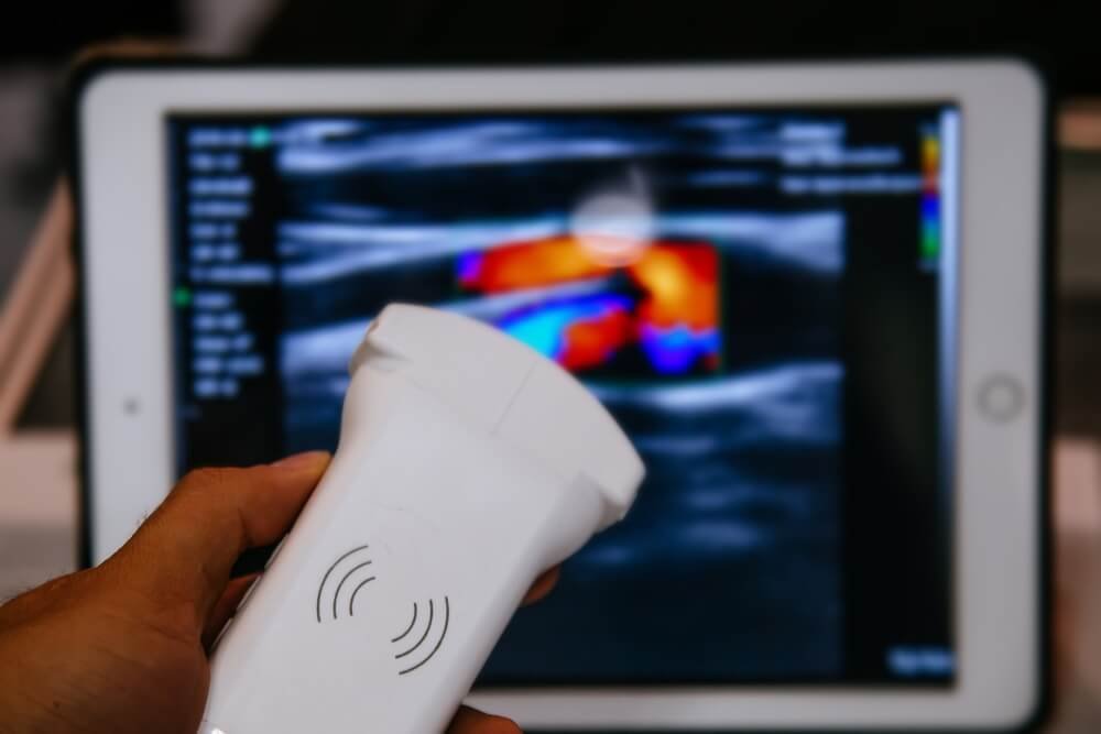
Portable ultrasound machines operate on the principle of sound wave reflection.
- High-frequency sound waves: These waves penetrate the skin and reflect off internal structures, creating images on the screen.
- Doppler imaging: This feature detects and displays blood flow within vessels, enabling differentiation between veins and arteries. The real-time visualization helps healthcare professionals assess vein size, depth, and patency, ensuring precise targeting.
How Portable Ultrasound Machines Help Find Veins
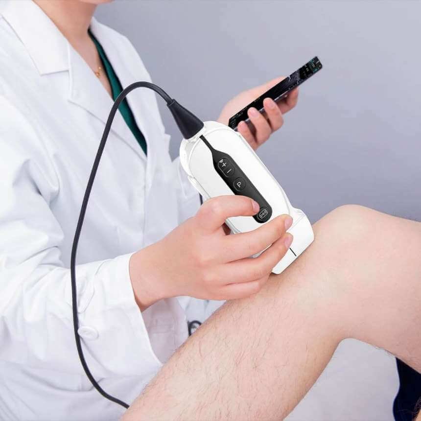
Portable ultrasound machines have revolutionized vein localization, making it easier, safer, and more efficient for medical professionals to locate veins, especially in challenging cases. Here’s an in-depth explanation of how these devices assist in vein finding:
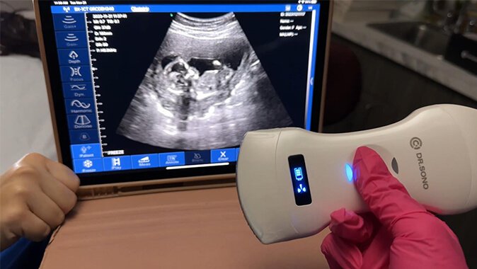
1. High-Frequency Sound Waves
Portable ultrasound machines use high-frequency sound waves to produce real-time images of the body’s internal structures, including veins. These sound waves are emitted by a transducer (probe) and reflected back when they encounter different tissues. The machine interprets these echoes to create detailed, dynamic images.
- Veins Appear as Dark Structures: In the ultrasound image, veins are usually depicted as compressible, dark (hypoechoic) structures.
- Clear Visualization: The machine allows medical professionals to see the veins’ size, depth, and course clearly.
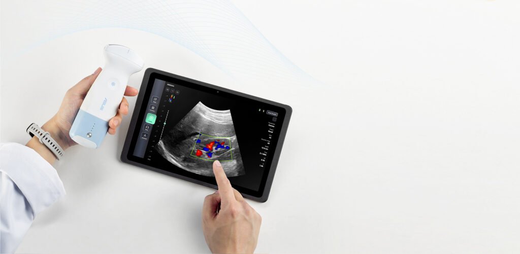
2. Doppler Imaging
Doppler imaging, a key feature in portable ultrasound machines, evaluates blood flow within the veins. This capability is crucial for distinguishing veins from arteries.
- Non-Pulsatile Flow: Veins exhibit a steady, non-pulsatile blood flow in Doppler mode.
- Enhanced Accuracy: By confirming blood flow direction, Doppler imaging ensures the identified structure is a vein and not an artery or another tissue.
3. Enhanced Imaging for Difficult Cases
Portable ultrasound machines are especially beneficial in situations where vein localization is challenging, such as:
- Small or Deep Veins: Ultrasound overcomes the limitations of visual or palpation methods, ensuring accurate targeting of veins that are not visible or palpable.
- Complicated Patient Profiles: Patients with conditions like obesity, edema, or scarring benefit from the detailed imaging provided by ultrasound.
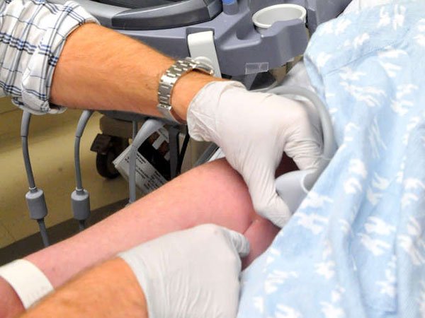
4. Real-Time Guidance
These machines provide real-time imaging, allowing healthcare providers to monitor their needle or catheter’s progress as it approaches the vein. This reduces the risk of:
- Missed Veins: Ensuring the needle enters the vein precisely.
- Complications: Avoiding accidental puncture of arteries, nerves, or surrounding tissues.
Benefits of Using a Portable Ultrasound Machine to Find a Vein
- Improved Accuracy
Ultrasound-guided vein localization minimizes guesswork. Clinicians can confidently access the vein on the first attempt, significantly reducing the risk of:- Missed veins
- Damage to surrounding tissues
- Reduced Complications
Repeated attempts at vein access can lead to:- Nerve damage: Accidentally striking nerves can result in pain or long-term issues.
- Hematoma formation: Puncturing adjacent tissues may cause blood pooling under the skin.
- Other complications: Infections and scarring are less likely when using ultrasound guidance.
- Enhanced Patient Safety
Ultrasound reduces unnecessary needle insertions, which:- Lowers the risk of introducing pathogens.
- Improves patient comfort and confidence in the procedure.
- Increased Efficiency
By improving the accuracy of vein localization, ultrasound-guided procedures are faster. This reduces:- Procedure time
- The need for repeat attempts, freeing up valuable time for both patients and clinicians.
Limitations and Challenges
- Operator Dependency
- The success of ultrasound-guided vein localization heavily relies on the skill of the operator. Inadequately trained users may misinterpret images, leading to errors.
- Regular training and practice are essential for mastering the technique.
- Image Quality
- Factors affecting image quality include the machine’s resolution, probe type, and operator handling. Older or basic models may not provide the clarity required for precise vein localization.
- The probe must be appropriately chosen and positioned to optimize visualization.
- Vein Depth and Size
- Deep veins or veins with small diameters may not be easily visualized, even with advanced equipment.
- Specialized probes, such as linear probes for shallow veins, may improve outcomes but require knowledge of their use.
- Patient Factors
- Obesity: Excess fat can obscure the veins, requiring advanced imaging techniques or higher-frequency probes.
- Edema: Fluid retention can distort images, making veins harder to locate.
- Abnormal anatomy: Congenital or acquired structural variations may present additional challenges.
Tips and Best Practices for Using a Portable Ultrasound Machine to Find a Vein
- Machine Selection
- Opt for machines with high-resolution imaging and Doppler capabilities.
- Ensure the device is portable, durable, and suitable for the clinical environment.
- Probe Selection
- Use a high-frequency linear probe for superficial veins.
- For deeper veins, a lower-frequency probe may be necessary.
- Scanning Technique
- Apply an adequate amount of ultrasound gel to ensure proper contact between the probe and skin.
- Use short, methodical sweeps to locate the vein, keeping the transducer at the appropriate angle for clear visualization.
- Image Optimization
- Adjust depth, gain, and focus settings to enhance clarity.
- Ensure the vein appears round (cross-section view) or tubular (longitudinal view) to confirm proper alignment.
Conclusion
Portable ultrasound machines are a game-changer in medical procedures requiring precise vein access. They provide unparalleled accuracy, safety, and efficiency, reducing complications and enhancing patient outcomes.
Despite certain limitations, such as operator dependency and challenges with image quality, these can be mitigated through training and proper equipment selection. As technology advances, portable ultrasound machines are becoming an integral part of modern medical practice, promising better care and more efficient procedures.
For healthcare professionals, investing in training and high-quality devices is a key to maximizing the benefits of this technology. Further research and development will only continue to improve its utility, ensuring even better outcomes for patients.

Article by
Scott Caswell
Scott is a co-founder of PUM and an ultrasound technology expert with a passion for innovation in the medical field. Scott has dedicated his career to advancing portable ultrasound devices, making medical imaging more accessible to professionals around the globe.
When not refining ultrasound devices, he enjoys hiking, experimenting with new recipes, and exploring the latest tech gadgets. Scott is dedicated to making healthcare more accessible and efficient through cutting-edge ultrasound solutions.
Join Our Mailing List & Save!
Enter your email address below to receive exclusive promotions and discounts along with additional product information and tips
Shop Now
Leave a Reply